See-Mode Breast & Thyroid
Automatically identifies and classifies nodules and lesions found in ultrasound imaging, aimed to reduce documentation and reporting time.

Overview
Thyroid nodules are characterized using the ACR TI-RADS and breast lesions are characterized by the BI-RADS guidelines. Following image acquisition by the ultrasound technologist, See-Mode analyzes the images, and then generates a preliminary worksheet for the ultrasound technologist to review. Worksheets along with radiologist impressions automatically flow through to PowerScribe/Fluency. See-Mode works with images captured using the standard clinical protocols for thyroid and breast ultrasound, and supports all major modality manufacturers.


Features
- Detection and image grouping: Automatic localization of lesion and nodule boundaries within thyroid ultrasound images, without the user having to indicate a region of interest. See-Mode can also automatically identify each annotated image in which a given lesion appears.
- Lesion characterization: For each identified lesion, it provides a full breakdown of the lesion features using BI-RADS is provided.
- Nodule characterization: For each identified nodule, it provides a full breakdown of the nodule features using TI-RADS is provided.
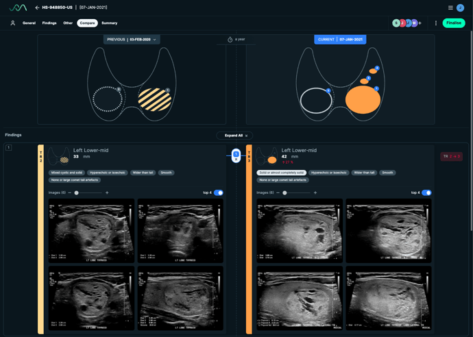
- Worksheet generation: Provides a digital worksheet, which can be reviewed and edited by the ultrasound technologist, along with a final PDF version which can be saved to the clinic’s PACS system. The worksheet includes a findings breakdown (with accompanying images), along with a diagram illustrating the lesion locations.
- Generates preliminary impressions which will be sent through to the radiologist reporting system. These impressions can then be reviewed/edited by the radiologist as required.
- Follow-up examinations: Enables matching lesion images from the current examination to images captured in a previous examination. Automatically compares new and old images, and output any identified changes in size or features.
Benefits
See-Mode aims to help clinicians by:
- Reducing repetitive and time-consuming tasks such as image analysis, measurement extraction, and report generation.
- Leveraging advanced pattern recognition and machine learning techniques to improve diagnostic accuracy, and reduce the chance of misdiagnosis.
- Standardizing and improving consistency and reporting across all examinations, regardless of the tech and interpreting radiologist.
Image examples


To find out more about this solution or any of the other 140+ applications on the Blackford Platform, please book a discovery call with our team.

Book a meeting
We’d welcome the opportunity to learn more about your AI needs and to explain how partnering with Blackford can drive efficiency and provide ongoing value.
Book a Meeting


.jpg)


