IB Delta T1 Maps
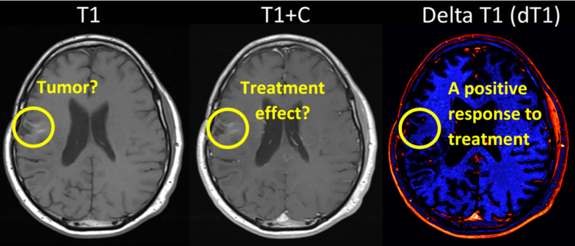
Comparison of standardized (calibrated) pre- (T1) and post-contrast (T1+C) anatomic images.

Overview
Reader misinterpretation and disagreement can contribute to radiologist burnout and diagnostic errors. Standard T1+C imaging can be variable and confounded by other bright enhancing signals, such as post-surgical blood product and may miss subtle enhancing regions which may not appear as an area of concern until weeks or months later in the treatment plan.
Delta T1 Maps aims to resolve this by detecting subtle regions of enhancing while subtracting out blood product and other artifacts that may appear bright on pre-contrast images.

Features
-
The IB Delta T1 module performs post-processing of DICOM images and simplifies post processing tasks.
-
It includes automated image registration features requiring minimal user interaction.
- It allows for the creation of difference images to ease identification of changes between two images (e.g., signal changes due to contrast uptake between pre- and post-contrast images).
Benefits
- Delta T1 maps aims to make the delineation of contrast enhancing regions more objective, faster and consistent.
- Delta T1 aims to predict early progression comparable with the standard 2d manual method but offers the potential for substantial time and cost savings for clinical trials.
Image examples


To find out more about this solution or any of the other 140+ applications on the Blackford Platform, please book a discovery call with our team.

Book a meeting
We’d welcome the opportunity to learn more about your AI needs and to explain how partnering with Blackford can drive efficiency and provide ongoing value.
Book a Meeting¹Schmainda KM, Prah MA, Zhang Z, Snyder BS, Rand SD, Jensen TR, et al. Quantitative Delta T1 (dT1) as a Replacement for Adjudicated Central Reader Analysis of Contrast-Enhancing Tumor Burden: A Subanalysis of the American College of Radiology Imaging Network 6677/Radiation Therapy Oncology Group 0625 Multicenter Brain Tumor Trial. AJNR American journal of neuroradiology [Internet]. 2019 Jul 1 [cited 2023 May 2];40(7):1132–9.


.jpg)


